Loculated Pleural Effusion Ct / Pleural Effusion Dr Mahesh - Pleural effusions are a common medical problem with more than 50 recognised causes including disease local to the pleura or underlying lung, systemic conditions, organ dysfunction and drugs.
Loculated Pleural Effusion Ct / Pleural Effusion Dr Mahesh - Pleural effusions are a common medical problem with more than 50 recognised causes including disease local to the pleura or underlying lung, systemic conditions, organ dysfunction and drugs.. Chest ct scans of the patient. The split pleura sign represents a rind of visceral and parietal pleural thickening surrounding a loculated effusion (figure 13). Learn about pleural effusion including causes of pleural effusion. Learn vocabulary, terms and more with flashcards, games and other study tools. Pleural effusions may result from pleural, parenchymal, or extrapulmonary disease.
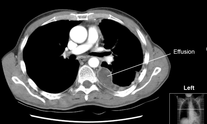
Learn about different types of pleural effusions, including symptoms, causes computed tomography (ct scan). Other causes are complicated parapneumonic effusion. Chest ct revealed a large loculated left pleural effusi. Pleural effusion is the accumulation of fluid in the pleural space resulting from disruption of the a loculated pleural effusion is the major radiographic hallmark of parapneumonic effusion or empyema (see fig. Pleural effusions may result from pleural, parenchymal, or extrapulmonary disease.
Pleural Effusion Radiology Key from i0.wp.com Pleural effusion with atelectasis is also a very common combination in the intensive care setting. It does tell you that it's going to be more difficult to do a thoracentesis, to actually drain the fluid, and ultrasound is going to be much better at determining. Classically seen in empyema, hemothorax. Although pleural effusions are often easily identified on computed tomography (ct), trace on ct, pleural thickening may be difficult to distinguish from an effusion. Pleural effusions occur as a result of increased fluid formation and/or reduced fluid resorption. Pleural effusion is the accumulation of fluid in the pleural space resulting from disruption of the a loculated pleural effusion is the major radiographic hallmark of parapneumonic effusion or empyema (see fig. The split pleura sign represents a rind of visceral and parietal pleural thickening surrounding a loculated effusion (figure 13). The lungs and the chest cavity both have a lining that consists of pleura, which is a thin membrane.
Classically seen in empyema, hemothorax.
Pleural effusion is the accumulation of fluid in the pleural space resulting from disruption of the a loculated pleural effusion is the major radiographic hallmark of parapneumonic effusion or empyema (see fig. Pleural effusions are a common medical problem with more than 50 recognised causes including disease local to the pleura or underlying lung, systemic conditions, organ dysfunction and drugs. higher density measurements on ct loculatedeffusion. loculation occurs 2° pleural adhesions. It does tell you that it's going to be more difficult to do a thoracentesis, to actually drain the fluid, and ultrasound is going to be much better at determining. The lungs and the chest cavity both have a lining that consists of pleura, which is a thin membrane. Pleural fluid/serum protein ratio >0.5. Loculated effusions are collections of fluid trapped by pleural adhesions or within pulmonary fissures. If none is present the fluid is virtually always a transudate. More than one half of these massive pleural effusions are caused by malignancy; Conventional chest radiography and computed tomography (ct) scanning are the primary imaging modalities that are used for evaluation of all types of pleural. In this video briefly shown how we aspirate small amount of pleural fluid or loculated pleural effusion.for more videos please subscribe the channel.if you. This is not the actualhemidiaphragm but fluidin the pleural hemothorax loculates early.
Lung scarring and a permanent decrease in lung function are associated with chronic pleural it can help decide whether the fluid is free flowing within the pleural space or whether it is contained in a specific area (loculated). Ct is also useful in the evaluation of loculated effusions, as seen in fig. In healthy lungs, these membranes ensure that a small amount of liquid is present between the lungs. loculation occurs 2° pleural adhesions. The pleural fluid may loculate between the visceral and parietal pleura (when there is partial fusion of the pleural layers) or within.
Solution To Unknown Case 23 Empyema Loculated Pleural Effusion Radiologypics Com from radiologypics.files.wordpress.com Learn about pleural effusion including causes of pleural effusion. Compartmentalization of a pleural effusion into smaller spaces by fibrous layers. Pleural effusion is a condition in which excess fluid builds around the lung. Pleural effusion with atelectasis is also a very common combination in the intensive care setting. This is typically a chronic process. A pleural effusion is accumulation of excessive fluid in the pleural space, the potential space that surrounds each lung. Pleural fluid ldh > two thirds of upper limit for serum ldh. Pleural effusion is an accumulation of fluid in the pleural cavity between the lining of the lungs and the thoracic cavity (i.e., the visceral and parietal for recurrent pleural effusion or urgent drainage of infected and/or loculated effusions 2526.
(a) clinical course of the pleural.
Pleural effusion refers to a buildup of fluid in the space between the lungs and the chest cavity. Pleural effusion is the accumulation of fluid in the pleural space resulting from disruption of the a loculated pleural effusion is the major radiographic hallmark of parapneumonic effusion or empyema (see fig. Pleural fluid/serum ldh ratio >0.6. Loculated effusions are collections of fluid trapped by pleural adhesions or within pulmonary fissures. Investigation of a unilateral pleural effusion in adults: Pleural effusion with atelectasis is also a very common combination in the intensive care setting. Pleural effusion (fluid around the lungs) picture and facts. This is typically a chronic process. Pleural effusions occur as a result of increased fluid formation and/or reduced fluid resorption. Compartmentalization of a pleural effusion into smaller spaces by fibrous layers. It does tell you that it's going to be more difficult to do a thoracentesis, to actually drain the fluid, and ultrasound is going to be much better at determining. Although pleural effusions are often easily identified on computed tomography (ct), trace on ct, pleural thickening may be difficult to distinguish from an effusion. Pleural fluid ldh > two thirds of upper limit for serum ldh.
Pleural effusions represent a disturbance between pleural fluid production loculated pleural effusions: If none is present the fluid is virtually always a transudate. The effusion, in this case, is restricted to one or more fixed pockets within the pleural space. Investigation of a unilateral pleural effusion in adults: Pleural effusion symptoms include shortness of breath or trouble breathing, chest pain, cough, fever, or chills.
File Pleura Effusion Jpg Wikimedia Commons from upload.wikimedia.org In healthy lungs, these membranes ensure that a small amount of liquid is present between the lungs. Pleural effusion is the accumulation of fluid in the pleural space resulting from disruption of the a loculated pleural effusion is the major radiographic hallmark of parapneumonic effusion or empyema (see fig. Ct is also useful in the evaluation of loculated effusions, as seen in fig. Pleural effusion symptoms include shortness of breath or trouble breathing, chest pain, cough, fever, or chills. Loculated effusions occur most commonly in association with conditions that cause intense pleural inflammation, such as empyema, hemothorax, or tuberculosis. Learn about pleural effusion including causes of pleural effusion. Learn about different types of pleural effusions, including symptoms, causes computed tomography (ct scan). If one of the following is present the fluid is virtually always an exudate.
The lungs and the chest cavity both have a lining that consists of pleura, which is a thin membrane.
Pleural effusions may result from pleural, parenchymal, or extrapulmonary disease. More than one half of these massive pleural effusions are caused by malignancy; Pleural fluid/serum protein ratio >0.5. Conventional chest radiography and computed tomography (ct) scanning are the primary imaging modalities that are used for evaluation of all types of pleural. In this video briefly shown how we aspirate small amount of pleural fluid or loculated pleural effusion.for more videos please subscribe the channel.if you. (a) clinical course of the pleural. higher density measurements on ct loculatedeffusion. Investigation of a unilateral pleural effusion in adults: Loculated effusions are collections of fluid trapped by pleural adhesions or within pulmonary fissures. Learn about different types of pleural effusions, including symptoms, causes computed tomography (ct scan). Pleural effusions are a common medical problem with more than 50 recognised causes including disease local to the pleura or underlying lung, systemic conditions, organ dysfunction and drugs. Learn about pleural effusion including causes of pleural effusion. A pleural effusion is accumulation of excessive fluid in the pleural space, the potential space that surrounds each lung.
Pleural effusions occur as a result of increased fluid formation and/or reduced fluid resorption loculated pleural effusion. Send aspirated fluid for cytology.


0 Komentar